2022
82. F. Zhong, S. Hu*, “A thin-film acoustic-optical combiner enables high-speed wide-field multi-parametric photoacoustic microscopy in reflection mode,” Optics Letters, (accepted).
81. A. Drieu, S. Du, S.E. Storck, J. Rustenhoven, Z. Papadopoulos, T. Dykstra, F. Zhong, K. Kim, S. Blackburn, T. Mamuladze, O. Harari, C.M. Karch, R.J. Bateman, R. Perrin, M. Farlow, J. Chhatwal, Dominantly Inherited Alzheimer Network, S. Hu, G. J. Randolph, I. Smirnov, and J. Kipnis, “Parenchymal border macrophages regulate the flow dynamics of the cerebrospinal fluid.” Nature (2022): 1-9. https://doi.org/10.1038/s41586-022-05397-3
80. Y. Zhou, N. Sun, and S. Hu, “Deep Learning-powered Bessel-beam Multi-parametric Photoacoustic Microscopy,” IEEE Transactions on Medical Imaging (2022). https://doi.org/10.1109/tmi.2022.3188739
79. A. Norambuena, X. Sun, H. Wallrabe, R. Cao, N. Sun, E. Pardo, N. Shivange, D.B. Wang, L.A. Post, H. A. Ferris, S. Hu, A. Periasamy, and G.S. Bloom, “SOD1 Mediates Lysosome-to-Mitochondria Communication and its Dysregulation by Amyloid-β Oligomers,” Neurobiology of Disease, 169 (2022): 105737. https://doi.org/10.1016/j.nbd.2022.105737
78. N. Sun, A.C. Bruce, B. Ning, R. Cao, Y. Wang, F. Zhong, S.M. Peirce, and S. Hu, “Photoacoustic Microscopy of Vascular Adaptation and Tissue Oxygen Metabolism during Cutaneous Wound Healing,” Biomedical Optics Express, 13, no. 5 (2022):2695-2706. (Editor’s pick) https://doi.org/10.1364/BOE.456198
77. Y. Wang, F. Zhong, N. Sun, Z. Xu, J. Li, Q. Liu, Z. Li, Z. Zuo, S. Hu, “High-speed Multi-parametric Photoacoustic Microscopy of Cerebral Hemodynamic and Metabolic Responses to Acute Hemodilution,” Optics Letters 47, no. 8 (2022): 1988-1991. https://doi.org/10.1364/OL.444327
76. A.B. Becker, L. Chen, B. Ning, S. Hu, J.A. Hossack, A.L. Klibanov, B.H. Annex, B.A. French, “Contrast-enhanced ultrasound reveals partial perfusion recovery after hindlimb ischemia as opposed to full recovery by laser Doppler perfusion imaging,” Ultrasound in Medicine & Biology 48, no. 6 (2022):1058-1069. https://doi.org/10.1016/j.ultrasmedbio.2022.02.002
2021
75. Z. Wang, Y. Zhou, and S. Hu, “Sparse Coding-enabled Low-fluence Multi-parametric Photoacoustic Microscopy,” IEEE Transactions on Medical Imaging 41, no. 4 (2021): 805-814. doi: 10.1109/TMI.2021.3124124
74. SG. Sathyanarayana†, Z. Wang†, N. Sun, B. Ning, S. Hu*, JA. Hossack*, “Recovery of Blood Flow From Undersampled Photoacoustic Microscopy Data Using Sparse Modeling,” IEEE Transactions on Medical Imaging 41, no. 1 (2021): 103-120. (*corresponding author; †equal contribution). doi: 0.1109/TMI.2021.3104521
73. V. Sciortino, A. Tran, N. Sun, R. Cao, T. Sun, Y. Sun, P. Yan, F. Zhong, Y. Zhou, C. Kuan, JM. Lee, S. Hu, “Longitudinal cortex-wide monitoring of cerebral hemodynamics and oxygen metabolism in awake mice using multi-parametric photoacoustic microscopy,” Journal of Cerebral Blood Flow & Metabolism, 41, no. 12 (2021): 3187-3199. https://doi.org/10.1177/0271678X211034096
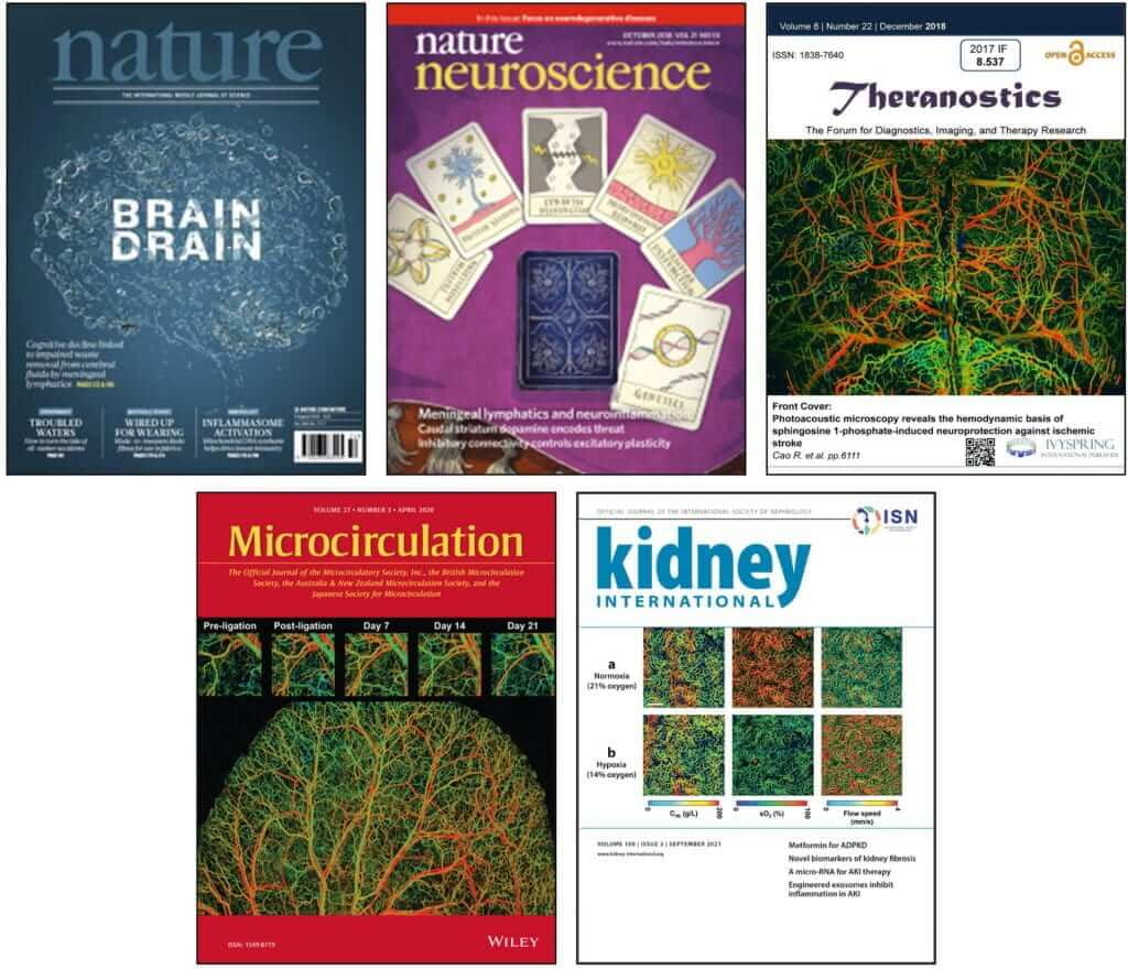
72. N. Sun, S. Zheng, D. Rosin, N. Poudel, J. Yao, H. Perry, R. Cao, M. Okusa, S. Hu, “Development of a photoacoustic microscopy technique to assess peritubular capillary function and oxygen metabolism in the mouse kidney,” Kidney International 100.3 (2021): 613-620. (Featured on cover) https://doi.org/10.1016/j.kint.2021.06.018
71. Y. Zhou, F. Zhong, S. Hu, “Temporal and spectral unmixing of photoacoustic signals by deep learning,” Optics Letters, 46, 2690-2693 (2021). https://doi.org/10.1364/OL.426678
70. Y. Zhou, F. Zhong, JM. Lee, S. Hu, “Simultaneous imaging of amyloid deposition and cerebrovascular function using dual-contrast photoacoustic microscopy,” Optics Letters. 46, 2561-2564 (2021). https://doi.org/10.1364/OL.419817
69. R. Cao, A. Tran, J. Li, Z. Xu, N. Sun, Z. Zuo, S. Hu, “Hemodynamic and Oxygen-metabolic Responses of the Awake Mouse Brain to Hypercapnia Revealed by Multi-parametric Photoacoustic Microscopy,” Journal of Cerebral Blood Flow and Metabolism, 41, no. 10 (2021): 2628-2639. https://doi.org/10.1177/0271678X211010352
2020
68. N. Poudel, S. Zheng, CM. Schinderle, N. Sun, S. Hu, MD. Okusa, “Peritubular Capillary Oxygen Consumption in Sepsis-Induced AKI: Multi-Parametric Photoacoustic Microscopy,” Nephron, 144(12):621-625 (2020). https://doi.org/10.1159/000511167
67. PS. Cottler, N. Sun, JM. Thuman, K. Bielak, L. Salopek, A. Piñeros-Fernandez, S. Hu, CA. Campbell, “The bio integration of a porcine acellular dermal matrix in a novel radiated breast reconstruction model”, Annals of Plastic Surgery, 84:S417-S423 (2020). doi: 10.1097/SAP.0000000000002277
66. F. Zhong, Y. Bao, R. Chen, Q. Zhou, S. Hu, “High-speed wide-field multi-parametric photoacoustic microscopy,” Optics Letters, 45(10), 2756-2759 (2020). https://doi.org/10.1364/OL.391824
65. N. Sun, B. Ning, A. Bruce, R. Cao, S. Seaman, T. Wang, R. Fritsche-Danielson, L. Carlsson, S. Peirce, S. Hu, “In vivo imaging of hemodynamic redistribution and arteriogenesis across a microvascular network,” Microcirculation, e12598 (2020). (Featured on cover) https://doi.org/10.1111/micc.12598
2019
64. Z. Xu, N. Sun, R. Cao, Z. Li, Q. Liu, S. Hu. “Cortex-wide multi-parametric photoacoustic microscopy based on real-time contour scanning,” Neurophotonics, 6, no. 3(2019), 035012. https://doi.org/10.1117/1.NPh.6.3.035012
63. AL. Klibanov, S. Hu. “Monitoring Oxygenation Levels Deep in the Tumor Core: Noninvasive Imaging of Hypoxia, Now in Real-Time 3D,” Cancer Research, 79(18):4577-4579 (2019). https://doi.org/10.1158/0008-5472.CAN-19-2151
62. Z. Xu, Y. Wang, N. Sun, Z. Li, S. Hu, Q. Liu. “Parallel computing for quantitative blood flow imaging in photoacoustic microscopy,” Sensors, 19(18), 4000 (2019). https://doi.org/10.3390/s19184000
61. R. Cao, C. Zhang, V. Mitkin, M. Lankford, J. Li, Z. Zuo, C. Meyer, C. Goyne, S. Ahlers, J. Stone, S. Hu. “Comprehensive characterization of cerebrovascular dysfunction in blast traumatic brain injury using photoacoustic microscopy,” Journal of Neurotrauma, 36(10):1526-1534 (2019). https://doi.org/10.1089/neu.2018.6062
60. PS. Cottler, JB. Olenczak, B. Ning, SA. Seaman, JM. Thuman, N. Sun, A. Piñeros-Fernandez, S. Hu, BR. DeGeorge Jr, CA. Campbell. “Fenestration improves acellular dermal matrix bio integration: an investigation of revascularization with photoacoustic microscopy”, Plastic and Reconstructive Surgery, 143(4):971-981 (2019).
doi: 10.1097/PRS.0000000000005410
59. R. Cao, J. Li, C. Zhang, Z. Zuo, S. Hu. “Photoacoustic microscopy of obesity-induced cerebrovascular alterations,” NeuroImage, 188:369-379 (2019). https://doi.org/10.1016/j.neuroimage.2018.12.027
58. T. Wang, N. Sun, R. Chen, Q. Zhou, S. Hu. “Isotropic-resolution photoacoustic microscopy with multi-angle illumination,” Optics Letters, 44(1), 1-4 (2019). https://doi.org/10.1364/OL.44.000001
2018
57. R. Cao, J. Li, Y. Kharel, C. Zhang, K. Lynch, Z. Zuo, S. Hu. “Photoacoustic microscopy of sphingosine 1-phosphate-induced neuroprotection against ischemic stroke,” Theranostics, 8(22), 6111-6120 (2018). doi: 0.7150/thno.29435
56. N. Sun, B. Ning, AC. Bruce, SA. Seaman, M. Rikard, CA. DeRosa, CL. Fraser, M. Wågberg, R. Fritsche-Danielson, J. Wikström, KR. Chien, A. Lundahl, M. Hölttä, K. Hansson, L. Carlsson, SM. Peirce, S. Hu. “Modified VEGF-A mRNA induces sustained multifaceted microvascular response and accelerates diabetic wound healing,” Scientific Reports, 8, 17509 (2018). https://doi.org/10.1038/s41598-018-35570-6
55. A. Norambuena, H. Wallrabe, R. Cao, DB. Wang, A. Silva, Z. Svindrych, S. Hu, RE. Tanzi, DY. Kim, GS. Bloom, “A novel lysosome-to-mitochondria signaling pathway disrupted by amyloid-β oligomers,” EMBO Journal, 37(22), e100241 (2018). https://doi.org/10.15252/embj.2018100241
54. A. Louveau, J. Herz, M. Alme, M. Dong, K. Viar, G. Herod, J. Knopp, J. Setliff, A. Lupi, S. Gabriel Ferreira Da Mesquita, E. Frost , R. Cao, S. Hu, J. Lukens, I. Smirnov, C. Overall, A. Salvador, G. Oliver, J. Kipnis. “CNS lymphatic drainage and neuroinflammation are regulated by meningeal lymphatic vasculature,” Nature Neuroscience, 21, 1380-1391 (2018). https://doi.org/10.1038/s41593-018-0227-9
53. S. Da Mesquita, A. Louveau, A. Vaccari, I. Smirnov, RC. Cornelison, KM. Kingsmore, C. Contarino, S. Onengut-Gumuscu, E. Farber, D. Raper, KE. Viar, RD. Powell, W. Baker, N. Dabhi, R. Cao, S. Hu, S. Rich, JM. Munson, CC. Overall, ST. Acton, J. Kipnis. “Functional aspects of meningeal lymphatics in aging and Alzheimer’s disease,” Nature 560, 185-191 (2018). https://doi.org/10.1038/s41586-018-0368-8
52. SG. Sathyanarayana, B. Ning, R. Cao, S. Hu, JA. Hossack. “Dictionary learning-based reverberation removal enables depth-resolved photoacoustic microscopy of cortical microvasculature in the mouse brain,” Scientific Reports, 8, 985 (2018). https://doi.org/10.1038/s41598-017-18860-3
51. H. Wallrabea, Z. Svindrych, SR. Alama, KH. Siller, T. Wang, D. Kashatus, S. Hu, A. Periasamy. “Segmented cell analyses to measure redox states of autofluorescent NAD(P)H, FAD & Trp in cancer cells by FLIM,” Scientific Reports, 8, 79 (2018). https://doi.org/10.1038/s41598-017-18634-x
2017
50. M. Gutknecht, M. Seaman, B. Ning, D. Cornejo, E. Mugler, P. Antkowiak, C. Moskaluk, S. Hu, FH. Epstein, KA. Kelly. “Identification of the S100 fused-type protein horner in as a regulator of tumor vascularity.” Nature Communications, 8(1), 552 (2017). https://doi.org/10.1038/s41467-017-00488-6
49. M. Kelly-Goss, B. Ning, A. Bruce, D. Tavakol, D. Yi, S. Hu, PA. Yates, SM. Peirce. “Dynamic, heterogeneous endothelial Tie2 expression and capillary blood flow during microvascular remodeling.” Scientific Reports, 7:9049 (2017). https://doi.org/10.1038/s41598-017-08982-z
48. R. Cao, J. Li, B. Ning, N. Sun, T. Wang, Z. Zuo, and S. Hu, “Functional and Oxygen-metabolic Photoacoustic Microscopy of the Awake Mouse Brain”, NeuroImage, 150: 77-87 (2017). https://doi.org/10.1016/j.neuroimage.2017.01.049
47. B. DeGeorge Jr., B. Ning, L. Salopek, A. Pineros-Fernandez, G. Rodeheaver, S. Peirce-Cottler, S. Hu, P. Cottler, C. Campbell, “Advanced imaging techniques for the investigation of acellular dermal matrix bio integration”, Plastic and Reconstructive Surgery, 139(2): 395-405 (2017). doi: 10.1097/PRS.0000000000002992
2016
46. T. Wang, N. Sun, R. Cao, B. Ning, R. Chen, Q. Zhou, and S. Hu, “Multi-parametric photoacoustic microscopy of the mouse brain with 300-kHz A-line rate”, Neurophotonics, 3(4), 045006 (2016). https://doi.org/10.1117/1.NPh.3.4.045006
45. R. Cao and S. Hu, “Molecular Photoacoustic Imaging of Energy Metabolism,” Molecular Imaging Gateway, 10(3) (2016). Read more
44. T. Keller, J. Butcher, G. Broseghini-Filho, C. Marziano, L. DeLalio, S. Rogers, B. Ning, J. Martin, S. Chechova, M. Cabot, X. Shu, A. Best, M. Good, A. Padilha, M. Purdy, M. Yeager, S. Peirce, S. Hu, A. Doctor, E. Barrett, T. Le, L. Columbus, and B. Isakson, “Modulating Vascular Hemodynamics With an Alpha Globin Mimetic Peptide (HbaX),” Hypertension, 68(6), 1494-1503 (2016). https://doi.org/10.1161/HYPERTENSIONAHA.116.08171
43. Y. Cho, G. Zheng, G. Augustine, D. Hochbaum, A. Cohen, T. Knöpfel, F. Pisanello, F. Pavone, I. Vellekoop, M. Booth, S. Hu, J. Zhu, Z. Chen, and Y. Hoshi “Roadmap on neurophotonics,” Journal of Optics, 18(9), 093007 (2016). http://dx.doi.org/10.1088/2040-8978/18/9/093007
42. J. Song, F. Wang, X. Yang, B. Ning, Harp,G. M. Culp, S. Hu, P. Huang, L. Nie, J. Chen, and X. Chen, “Gold nanoparticle coated carbon nanotube ring with enhanced Raman scattering and photothermal conversion property for theranostic applications,” Journal of the American Chemical Society, 138(22), 7005-7015 (2016). https://doi.org/10.1021/jacs.5b13475
41. S. Hu, “Emerging concepts in functional and molecular photoacoustic imaging,” Current Opinion in Chemical Biology, 33, 25-31(2016). https://doi.org/10.1016/j.cbpa.2016.04.003
40. C. Yeh, J. Liang, Y. Zhou, S. Hu, R. E. Sohn, J. M. Arbeit, and L. V. Wang, “Photoacoustic microscopy of arteriovenous shunts and blood diffusion in early-stage tumors,” Journal of Biomedical Optics, 21(2), 020501-020501 (2016). https://doi.org/10.1117/1.JBO.21.2.020501
39. S. Hu, “Listening to the brain with photoacoustics,” IEEE Journal of Selected Topics in Quantum Electronics, 22(3), 6800610 (2016). doi: 10.1109/JSTQE.2015.2487890
2015
38. B. Ning, N. Sun, R. Cao, R. Chen, K. K. Shung, J. A. Hossack, J.-M. Lee, Q. Zhou, and S. Hu, “Ultrasound-aided multi-parametric photoacoustic microscopy of the Mouse Brain,” Scientific Reports, 5, 18775 (2015). https://doi.org/10.1038/srep18775
37. K. Song, P. Huang, C. Yi, B. Ning, S. Hu, L. Nie, X. Chen, and Z. Nie, “Photoacoustic and Colorimetric Visualization of Latent Fingerprints,” ACS Nano, 9(12), DOI: 10.1021/acsnano.5b05618 (2015). https://doi.org/10.1021/acsnano.5b05629
36. T. Wang, R. Cao, B. Ning, A. J. Dixon, J. A. Hossack, A. L. Klibanov, Q. Zhou, A. Wang, and S. Hu, “All-optical photoacoustic microscopy based on plasmonic detection of broadband ultrasound,” Applied Physics Letters. 107, 153702 (2015). https://doi.org/10.1063/1.4933333
35. A. J. Dixon, S. Hu, A. L. Klibanov, and J. A. Hossack, “Oscillatory Dynamics and In Vivo Photoacoustic Imaging Performance of Plasmonic Nanoparticle-Coated Microbubbles,” Small, 11(25), 3066–3077 (2015). https://doi.org/10.1002/smll.201403398
34. R. Cao, J. P. Kilroy, B. Ning, T. Wang, J. A. Hossack, and S. Hu, “Multispectral photoacoustic microscopy based on an optical-acoustic objective,” Photoacoustics, 3(2), 55-59 (2015). https://doi.org/10.1016/j.pacs.2014.12.004
33. B. Ning, M. J. Kennedy, A. J. Dixon, N. Sun, R. Cao, B. T. Soetikno, R. Chen, Q. Zhou, K. K. Shung, J. A. Hossack, and S. Hu, “Simultaneous photoacoustic microscopy of microvascular anatomy, oxygen saturation, and blood flow,” Optics Letters, 40(6), 910-913 (2015). https://doi.org/10.1364/OL.40.000910
2014
32. L. Li, C. Yeh, S. Hu, L. Wang, B. T. Soetikno, R. Chen, Q. Zhou, K. K. Shung, K. I. Maslov, and L. V. Wang, “Fully motorized optical-resolution photoacoustic microscopy,” Opt. Lett. 39, 2117-2120 (2014). https://doi.org/10.1364/OL.39.002117
31.Y. Zhou, X. Yi, W. Xing, S. Hu, K. Maslov, and L. V. Wang, “Microcirculatory changes identified by photoacoustic microscopy in patients with complex regional pain syndrome type I after stellate ganglion blocks,” J. Biomed. Opt. 19(8), 086017 (2014). https://doi.org/10.1117/1.JBO.19.8.086017
30. C. Yeh, B. Soetikno, S. Hu, K. I. Maslov, and L. V. Wang, “Microvascular quantification based on contour-scanning photoacoustic microscopy,” J. Biomed. Opt. 19(9), 096011 (2014). https://doi.org/10.1117/1.JBO.19.9.096011
29. C. Yeh, B. Soetikno, S. Hu, K. I. Maslov, and L. V. Wang, “Three-dimensional arbitrary trajectory scanning photoacoustic microscopy,” J. Biophoton., 201400055 (2014). https://doi.org/10.1002/jbio.201400055
Pre-2013
28. S. Hu, and L. V. Wang. “Optical-resolution photoacoustic microscopy: auscultation of biological systems at the cellular level,” Biophysical journal 105, no. 4 (2013): 841-847. https://doi.org/10.1016/j.bpj.2013.07.017
27.L. V. Wang and S. Hu, “Photoacoustic tomography is ready to revolutionize,” BioOptics World 6, 32–37 (2013). (Featured on the cover) Read more
26. C. Yeh; S. Hu; K. Maslov, and L. V. Wang, “Photoacoustic microscopy of blood pulse wave”, Journal of Biomedical Optics 17(7), 070504 (2012). https://doi.org/10.1117/1.JBO.17.7.070504
25. L. V. Wang and S. Hu, “Photoacoustic tomography: in vivo imaging from organelles to organs,” Science 335(6075), 1458-1462 (2012). doi: 10.1126/science.1216210
24. P. K. Avti, S. Hu, C. P. Favazza, A. G. Mikos, J. A. Jansen, K. R. Shroyer, L. V. Wang, and B. Sitharaman, “Detection, mapping, and quantification of single-walled carbon nanotubes in histological specimens with photoacoustic microscopy,” PLoS One 7(4), e35064 (2012). https://doi.org/10.1371/journal.pone.0035064
23. C. Zhang, K. Maslov, S. Hu, R. Chen, Q. Zhou, K. K. Shung, and L. V. Wang, “Reflection-mode submicron-resolution in vivo photoacoustic microscopy,” Journal of Biomedical Optics 17(2), 020501 (2012). https://doi.org/10.1117/1.JBO.17.2.020501
22. X. Cai, B. S. Paratala; S. Hu, B. Sitharaman, and L. V. Wang, “Multiscale Photoacoustic Microscopy of Single-Walled Carbon Nanotube-Incorporated Tissue Engineering Scaffolds,” Tissue Engineering Part C: Methods 18(4), doi:10.1089/ten.tec.2011.0519 (2012). https://doi.org/10.1089/ten.tec.2011.0519
21. S. Oladipupo, S. Hu, J. Kovalski, J. Yao, A. C. Sanford, R. Sohn, R. V. Shohet, K. Maslov, L. V. Wang, and J. M. Arbeit, “VEGF is essential for hypoxia-inducible factor-mediated neovascularization but dispensable for endothelial sprouting,” Proceedings of the National Academy of Sciences 108(32), 13264-13269 (2011). https://doi.org/10.1073/pnas.1101321108
20. V. Tsytsarev, S. Hu, J. Yao, K. Maslov, D. L. Barbour, and L. V. Wang, “Photoacoustic microscopy of microvascular responses to cortical electrical stimulation,” Journal of Biomedical Optics 16(7), 076002 (2011). (Selected for inclusion in Faculty of 1000) https://doi.org/10.1117/1.3594785
19. H.-W. Wang, N. Chai, P. Wang, S. Hu, W. Dou, D. Umulis, L. V. Wang, M. Sturek, R. Lucht, and J.-X. Cheng, “Label-free bond-selective imaging by listening to vibrationally excited molecules,” Physical Review Letters 106(23), 238106 (2011). (Highlighted by Science) https://doi.org/10.1103/PhysRevLett.106.238106
18. S. Hu, K. Maslov, and L. V. Wang, “Three-dimensional optical-resolution photoacoustic microscopy,” Journal of Visualized Experiments, DOI: 10.3791/2729 (2011). doi: 10.3791/2729
17. S. Oladipupo; S. Hu; A. C. Sanford, J. Yao, J. Kovalski, R. V. Shohet, K. Maslov, L. V. Wang, and J. M. Arbeit, “Conditional HIF-1 induction produces multistage neovascularization with stage-specific sensitivity to VEGFR inhibitors and myeloid cell independence,” Blood 117(15), 4142-4153 (2011). https://doi.org/10.1182/blood-2010-09-307538
16. S. Hu; K. Maslov; and L. V. Wang, “Second-generation optical-resolution photoacoustic microscopy with improved sensitivity and speed,” Optics Letters 36(7), 1134–1136 (2011). (Featured article) https://doi.org/10.1364/OL.36.001134
15. Y. Wang, S. Hu, K. Maslov, Y. Zhang, Y. Xia, and L. V. Wang, “In vivo integrated photoacoustic and confocal microscopy of hemoglobin oxygen saturation and oxygen partial pressure,” Optics Letters 36(7), 1029–1031 (2011). https://doi.org/10.1364/OL.36.001029
14. Y. Wang, K. Maslov, Y. Zhang, S. Hu, L. Yang, Y. Xia, J. Liu, L. V. Wang, “Fiber-laser-based photoacoustic microscopy and melanoma cell detection,” Journal of Biomedical Optics 16(1), 011014 (2011). https://doi.org/10.1117/1.3525643
13. Y. Wang, K. Maslov, C. Kim, S. Hu, and L. V. Wang, “Integrated photoacoustic and fluorescence confocal microscopy,” IEEE Transactions on Biomedical Engineering 57(10), 2576–2578 (2010). doi: 10.1109/TBME.2010.2059026
12. S. Hu and L. V. Wang, “Neurovascular photoacoustic tomography,” Frontiers in Neuroenergetics 2(10), doi:10.3389/fnene.2010.00010 (2010). https://doi.org/10.3389/fnene.2010.00010
11. Z. Guo, S. Hu, and L. V. Wang, “Calibration-free absolute quantification of optical absorption coefficients using acoustic spectra in three-dimensional photoacoustic microscopy of biological tissue,” Optics Letters 35(12), 2067–2069 (2010). https://doi.org/10.1364/OL.35.002067
10. S. Hu and L. V. Wang, “Photoacoustic imaging and characterization of the microvasculature,” Journal of Biomedical Optics 15(1), 011101 (2010). (4th most cited article in Journal of Biomedical Optics since 2009) https://doi.org/10.1117/1.3281673
9. S. Hu, B. Rao, K. Maslov, and L. V. Wang, “Label-free photoacoustic ophthalmic angiography,” Optics Letters 35(1), 1–3 (2010). (Featured by Virtual Journal of Biomedical Optics) https://doi.org/10.1364/OL.35.000001
8. S. Hu; P. Yan; K. Maslov, J.-M. Lee, and L. V. Wang, “Intravital imaging of amyloid plaques in a transgenic mouse model using optical-resolution photoacoustic microscopy,” Optics Letters 34(24), 3899–3901 (2009). https://doi.org/10.1364/OL.34.003899
7. J. Yao, K. Maslov, S. Hu, and L. V. Wang, “Evans blue dye-enhanced capillary-resolution photoacoustic microscopy in vivo,” Journal of Biomedical Optics 14(5), 054049 (2009). https://doi.org/10.1117/1.3251044
6. S. Hu, K. Maslov, V. Tsytsarev, and L. V. Wang, “Functional transcranial brain imaging by optical-resolution photoacoustic microscopy,” Journal of Biomedical Optics 14(4), 040503 (2009). (6th most cited article in Journal of Biomedical Optics since 2009) https://doi.org/10.1117/1.3194136
5. S. Hu, K. Maslov, and L. V. Wang, “In vivo functional chronic imaging of a small animal model using optical-resolution photoacoustic microscopy,” Medical Physics 36(6), 2320-2323 (2009). https://doi.org/10.1118/1.3137572
4. S. Hu, K. Maslov, and L. V. Wang, “Noninvasive label-free imaging of microhemodynamics by optical-resolution photoacoustic microscopy,” Optics Express 17(9), 7688-7693 (2009). https://doi.org/10.1364/OE.17.007688
3. K. Maslov; H. F. Zhang; S. Hu; and L. V. Wang, “Optical-resolution photoacoustic microscopy for in vivo imaging of single capillaries,” Optics Letters 33(9), 929–931 (2008). (5th most cited article in Optics Letters since 2007) https://doi.org/10.1364/OL.33.000929
2. Y. Wang, S. Hu, L. Yan, J.-Y. Yang, and A. E. Willner, “Chromatic dispersion and polarization mode dispersion monitoring for multi-level intensity and phase modulation systems,” Optics Express 15(21), 14038-14043 (2007). https://doi.org/10.1364/OE.15.014038
1. S. Hu, H. Zhang, and Y. Guo, “Stiffness analysis in the numerical solution of Raman amplifier propagation equation,” Optics Express 12(8), 1656-1664 (2004). https://doi.org/10.1364/OPEX.12.001656